
Files
Archive Date
2015
Keywords
knee, biomechanics, laxity, in vitro, finite element
Disciplines
Biomechanics and Biotransport | Biomedical Engineering and Bioengineering
Recommended Citation
Harris MD, Cyr AJ, Ali AA, Fitzpatrick CK, Rullkoetter P, Maletsky L Shelburne KB. A combined experimental and computational approach to subject-specific analysis of knee joint laxity. J Biomech Eng. 2016. 138(8). Ali AA, Shalhoub SS, Cyr AJ, Fitzpatrick CK, Maletsky LP, Rullkoetter PJ, Shelburne KB, 2016. Validation of predicted patellofemoral mechanics in a finite element model of the healthy and cruciate-deficient knee. J Biomech 49:302-9.
Specimen Description
Age: 37 y/o; Sex: Female; Weight: 54.43 kg; Height: 168 cm; BMI: 19.37
CT Image Acquisition Details
NA
MR Image Acquisition Details
Sequence: T2 trufi3d_we_SAG; 192 slices; thickness: 0.5 mm; in-plane pixel spacing (mm): 0.46875 x 0.46875; in-plane resolution: 320 x 320
Document Type
Data Set
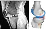
Kinematics and Load during Laxity Testing Data Details
We have provided kinematic and loading data for knee envelope (i.e. laxity) tests of intact knees and following resection of the cruciate ligaments. Please refer to and cite the following for details about the data collection process: Harris MD, Cyr AJ, Ali AA, Fitzpatrick CK, Rullkoetter P, Maletsky L, Shelburne KB. A combined experimental and computational approach to subjects-specific analysis of knee joint laxity. J Biomech Eng. 2016. 138(8).
For each knee, the kinematic and load data are subdivided by the experimental test being performed. For example, the initial sets of provided data were recorded during a passive knee envelope test with the knee intact (i.e. before resecting the cruciate ligaments or meniscus). Thus, the data are located in a folder called INTACT_KE for intact knee envelope.
Examples of the naming conventions for the Grood-Suntay kinematics and loads:
- DU02_INTACT_KE_AP.csv – These are kinematic and load data as the knee is being manually flexed between approximately 0 and 130 degrees and alternating anterior and posterior forces are being simultaneously applied. Although the anterior-posterior forces are predominant, we have supplied rotations, translation, forces, and torques in all 6 DOF.
- DU02_INTACT_KE_IE.csv – These are kinematic and load data as the knee is being manually flexed between approximately 0 and 130 degrees and alternating internal and external torques are being simultaneously applied.
- DU02_INTACT_KE_VV.csv – These are kinematic and load data as the knee is being manually flexed between approximately 0 and 130 degrees and alternating varus and valgus torques are being simultaneously applied.
- DU02_INTACT_KE_Passive.csv – These are kinematic and load data as the knee is being manually flexed between approximately 0 and 130 degrees.
In addition, Excel files are included processing of the experimental data that separated the response of the knee into three loading directions: varus-valgus (VV), internal-external (IE), and anterior-posterior (AP). The naming convention is for example: “DU02_Resected_experimental_processed”
3D Reconstruction Details
We have supplied subjects-specific reconstructions of the bone and cartilage geometry of the femur, tibia, fibula, femoral cartilage, and tibial cartilage for each of the cadaveric knees. The reconstruction files are in STL ASCII format. These reconstructions come directly from segmentation of the MR images and have not undergone the refinement and additional smoothing for finite element modeling. It is left to the user to use the reconstructions as desired.
An example of the naming convention for reconstruction files:
- DU02_Fem_Bone.stl – This is the femur bone reconstruction for knee # DU02
NOTE: On the DU01 knee, the bones were segmented from CT data and then aligned to the cartilage, which had been aligned from the MRI. All other knees were segmented entirely form the MRI.
Experimental Probe Points Details
For registration of bony structures during different testing protocols and for placement of soft-tissue meshes during finite element modeling, an Optotrak motion capture system and digitizing wand were used to probe several locations on the bone, cartilage, and ligament attachments. These probe points have been transformed to align with the 3D reconstructions and MR images.
The probed structures include: femur bone, tibia bone, articular geometry of the femur cartilage, tibia cartilage, and patella cartilage, and ligament attachment sites of the LCL, MCL, ACL, and PCL.
Examples of the naming convention for probed point files:
- DU02_ACL_Fem.txt – These are the probed ACL attachments on the femur for knee #DU02
- DU02_Fem_artgeo.txt – These are probed points on the articular cartilage surface of the femur for knee #DU02
Five points were digitized on bone (femur, tibia, patella), from which local coordinate systems were established. Using the local coordinate systems and data recorded from the Optotrak motion capture system data (i.e. from tracking bodies mounted to each bone), joint angles of the tibia with respect to the femur (TF), patella with respect to the femur (PF), and patella with respect to the tibia (PT) kinematics were calculated. The resulting kinematics and corresponding 6 DOF torques and forces, recorded by the load cell mounted on the tibia can be found in the Kinematics and Loads section.
DU02_Fem_GSPts.txt – These are the digitized points on the femur, used to establish a local coordinate system and calculate Grood-Suntay kinematics for the femur of knee #DU02
Publication Statement
License Agreement
Downloading any of the provided data indicates your compliance with the following license agreement.
USE AGREEMENT
Permission is hereby granted, free of charge, to any person obtaining a copy of this experimental, image, and computational model data, and associated documentation files (the “Data”) to be used without restriction to use, copy, merge, and publish the Data for non-commercial purposes, including but not limited to non-commercial research and non-profit teaching or learning. Permission is furnished subject to the following conditions: 1. Relevant publications by the University of Denver must be cited as the source the Data in all cases of publication or distribution. 2. Where applicable, University of Denver personnel will be included as co-authors in cases of publication. Use of the Data for any commercial application must be explicitly obtained by the University of Denver Center for Orthopaedic Biomechanics.
LIABILITY AGREEMENT
The Data is provided “as is” with no express or implied warranty or guarantee. The University of Denver and the Center for Orthopaedic Biomechanics do not accept any liability or provide any guarantee in connection with uses of the Data, including but not limited to, fitness for a particular purpose and noninfringement. The University of Denver and the Center for Orthopaedic Biomechanics are not liable for direct or indirect losses or damage, of any kind, which may arise through the use of this data.
CITATIONS
Knee Laxity Testing Data and Models: Harris MD, Cyr AJ, Ali AA, Fitzpatrick CK, Rullkoetter P, Maletsky L Shelburne KB. A combined experimental and computational approach to subjects-specific analysis of knee joint laxity. J Biomech Eng. 2016. 138(8). doi: 10.1115/1.4033882
Knee Extension Testing Data and Models: Ali AA, Shalhoub SS, Cyr AJ, Fitzpatrick CK, Maletsky LP, Rullkoetter PJ, Shelburne KB, 2016. Validation of predicted patellofemoral mechanics in a finite element model of the healthy and cruciate-deficient knee. J Biomech 49:302-9.