

Anatomical Variation in the Knee: A Dataset of 3D Bone Models and Statistical Shape Models
Preview
Files
Download Metadata and Morphological Parameters (769 KB)
Download Raw Segmentation Masks (192.9 MB)
Download Raw 3D Models (1548.2 MB)
Download Smoothed Segmentation Masks (192.5 MB)
Download Smoothed 3D Models (1502.0 MB)
Download Registered 3D Models (496.1 MB)
Download Registers (299.2 MB)
Download Visualization Code and Outputs (34.7 MB)
Creation Date
7-31-2026
Description
Objects Available for Download
- Metadata and Morphological Parameters (.xlsx) Size: 770 KB
- Raw Segmentation Masks (.MHD, .RAW) Zip size: 192 MB Extracted size: 166 GB
- Raw 3D Models (STL) Zip size: 1.51 GB Extracted size: 3.34 GB
- Smoothed Segmentation Masks (.MHD, .RAW) Zip size: 192 MB Extracted size: 166 GB
- Smoothed 3D Models (STL) Zip size: 1.46 GB Extracted size: 3.26 GB
- Registered 3D Models (STL, .inp) Zip size: 496 MB Extracted size: 1.42 GB
- Registers (.mat) Size: 299 MB
- Visualization Code and Outputs (.m) Size: 34.78 MB
Data Records
The dataset includes data and metadata for 102 subjects, 50 female and 52 male, with an average age of 45.3 (+/- 14.1) and 43.9 (+/- 13.9) years, respectively. Subjects came from Asian (A, n = 12), Non-Hispanic Black or African American (NHB, n = 23), Hispanic (H, n = 21), Native American (NA, n = 24), and Non-Hispanic White (NHW, n = 22) racial or ethnic backgrounds. NMDID race and ethnicity metrics were self-reported by the decedents in the 2010 census. Please note that for the purposes of this work, "Hispanic" refers to decedents who identified as racially Hispanic, reflecting the usage in the 2010 census where many New Mexicans selected Hispanic as an "Other" race option.
Each subject is identified by a unique six-digit NMDID code, enabling association with the original scan data. Access to the NMDID requires completion of the database’s data access, sharing, and usage agreement. For each subject, the dataset includes select metadata and morphologic parameters, raw and smoothed segmentation masks for the tibia, femur, and fibula of a single lower extremity; raw and smoothed segmented bony anatomy in CT space; and registered bony anatomy for the distal femur and proximal tibia.
- Metadata:
- Metadata are provided in the Excel file Metadata_and_MorphologicalParameters.xlsx under the ‘Metadata’ worksheet and are organized by NMDID identifier. The included data fields are sex, age (years), race, ethnicity, additional ancestry information, socioeconomic status (childhood), socioeconomic status (adulthood), living height (cm), living weight (kg), cadaver height (cm), BMI, and laterality of the segmented limb (left or right). Information on metadata collection protocols and metadata may be obtained through the NMDID.
- Morphological parameters:
- Calculated morphological parameters are provided in the same Excel file under the ‘Morphological Parameters’ worksheets and are organized by NMDID identifier. These parameters are also available in MATLAB within the Registers.mat structure under the field MP. Parameter definitions and reference figures illustrating the parameters are also included in the Excel file under the ‘Parameter Definitions’ worksheets.
- Raw Segmentation Masks:
- Raw segmentation masks of the proximal femur, distal femur, tibia, and distal fibula were exported from ScanIP as paired .MHD (MetaImage header) and .RAW (binary image data) files in the original CT coordinate space. These masks have not undergone any post-segmentation processing, such as smoothing. They are provided for users who wish to modify the segmentation prior to generating their own bone models or for applications that require segmentation masks rather than surface models. If desired, these files can be imported into software such as 3D Slicer (slicer.org) and converted to other formats (e.g. .NRRD or TIFF stacks). These masks are identified by the label ‘rawsegmask’.
- Raw 3D Models:
- Raw 3D models of the proximal femur, distal femur, tibia, and distal fibula were exported from ScanIP as STL files in the original CT coordinate space using the program’s default edge length (approximately 0.33 mm). These models have not undergone any smoothing and may include holes and/or self-intersections. They are intended for those who wish to perform their own post-segmentation processing and are denoted by the label ‘raw’ in the filename.
- Smoothed Segmentation Masks:
- Smoothed segmentation masks of the proximal femur, distal femur, tibia, and distal fibula are also provided. These masks were exported from ScanIP as .MHD/.raw file pairs in the original CT coordinate space and are identified by the label “smoothedsegmask.”
- Smoothed 3D Models:
- Smoothed 3D models are also provided for the proximal femur, distal femur, tibia, and distal fibula. Like the Raw 3D models, these were exported from ScanIP as STL files in the original CT coordinate space using the program’s default edge length (approximately 0.33 mm). These models are identified by the label ‘smoothed’.
- Registered 3D Models:
- Registered models of the distal tibia and proximal femur are provided as STL files in the final aligned (“register”) space. Each tibia model consists of 20,296 nodes and 40,588 triangular elements, while each femur model contains 49,767 nodes joined by 99,530 triangular elements. These models are identified by the label ‘registered’. For compatibility with finite element software, the registered models are also provided in Abaqus .inp format (Dassault Systems). These text-based files include the list of nodes (*NODE) and elements (*ELEMENT) defining the mesh connectivity. These files can be identified by the label ‘registeredsurf’.
- Registers:
- The surface SSM registrations are provided in a single .mat file (filename: Registers.mat) containing two structures corresponding to the distal femur (“Femur”) and proximal tibia (“Tibia”). Each structure includes element connectivity, the surface SSM registration matrix, registered nodal coordinates in column format, and the coordinate transformation matrices. Element connectivity is stored as an m × 4 matrix, where m is the number of triangular elements defining the registered surface. Each row of the matrix corresponds to one triangular surface element, with columns denoting the element number followed by the indices of the three nodes defining that element. The surface SSM register is provided as a .mat file containing a matrix of aligned registered nodal coordinates. Each column of the matrix corresponds to a single registered surface instance. The matrix is defined as R = [X1, X2, ..., X102] where Xi is a column vector representing the ith registered instance. Each Xi is constructed by concatenating the (x, y, z) coordinates of the n nodes into a vector of length 3n. This format allows nodal correspondence to be preserved across all instances. For convenience, the nodal coordinates for each registered instance are also provided separately in a column format, with each row in the matrix providing the x, y, and z coordinates for the node. All relevant coordinate transformation matrices are also included in this file to facilitate use of the registered bone models and reproduction of the SSMs. These include an overall transformation matrix converting coordinates from CT space to the aligned register space, as well as intermediate transformations from CT to anatomical space, anatomical to template space, and template to aligned register space.
The complete dataset consists of 4.17 GB of metadata and data. To facilitate downloading, the dataset has been organized into separate folders according to the data types described above. File names are structured to identify the subject's NMDID code, bone (femur or tibia), and processing state.
Code Availability
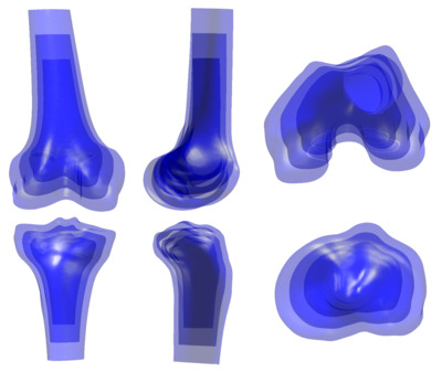
A MATLAB (MathWorks, Natick, MA) script is provided to facilitate visualization of the anatomy and SSMs (filename: SSMvisualization.m). It imports Registers.mat (described under ‘Registers’ in the Data Records section) and, after the user selects either the femur or tibia, constructs a statistical shape model using PCA. The code enables visualization of the modes of shape variation through two-dimensional overlays at ±2 standard deviations in multiple views, animated videos illustrating each mode in multiple views, and scatter plots of principal component scores for all subjects grouped by sex and race or ethnicity. All outputs are automatically saved in their appropriate formats to a folder labeled ‘outputs’.
This framework is intended to demonstrate how to interact with and visualize the statistical shape models and their instances. It is not intended to provide an exhaustive set of analysis or visualization tools, and users are encouraged to adapt and extend the framework to suit their own applications.
Contributing New and Updated Geometries
New geometries can be added to the dataset by contacting the authors ([email protected] and [email protected]). The authors will check new or revised content for accuracy and completeness and update the folders with credit of the contributors highlighted on this website.
Organizational Units
Daniel Felix Ritchie School of Engineering and Computer Science, Center for Orthopaedic Biomechanics
Copyright Statement / License for Reuse

This work is licensed under a Creative Commons Attribution-NonCommercial 4.0 International License
Publisher
University of Denver
Document Type
Data Set
City
Denver, CO
Publication Statement
License
Creative Commons License
This work is licensed under a Creative Commons Attribution Non-Commercial 4.0 International License
Liability Agreement
The Data is provided “as is” with no express or implied warranty or guarantee. The University of Denver and the Center for Orthopaedic Biomechanics do not accept any liability or provide any guarantee in connection with uses of the Data, including but not limited to, fitness for a particular purpose and noninfringement. The University of Denver and the Center for Orthopaedic Biomechanics are not liable for direct or indirect losses or damage, of any kind, which may arise through the use of this data.
Citations
This work has been submitted and is in review at the following journal: Kindy, G., Alsaadi, O., Laz, P.J. “Anatomical Variation in the Knee: A Dataset of 3D Bone Models and Statistical Shape Models.” Nature: Scientific Data, in review.