
Files
Download All (215 KB)
Archive Date
4-2019
Keywords
Knee, Biomechanics, In vivo, Kinematics, Patella, Tibia, Femur
Disciplines
Biomechanics | Biomechanics and Biotransport | Biomedical Engineering and Bioengineering | Kinesiology
Specimen Description
Subjects Gender Height (cm) Weight (Kg) Age BMI 1 Female 150 57 64 25.3 2 Female 163 65.8 56 24.8 3 Male 174 74.8 61 24.7 4 Female 143.5 72.6 57 35.5 5 Female 163 130 81 34.9 6 Male 181.6 88.9 70 27 7 Male 177.2 74.4 60 23.7 8 Male 170.2 71.1 66 24.6
Data Acquisition and Processing Methods
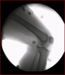
High speed stereo radiography (HSSR) was used to measure the kinematics of the TF and PF joints in each subject’s dominant knee. HSSR captures two radiographic views to enable three-dimensional tracking of the bones in the knee. The HSSR system is composed of two matching custom radiography systems with 40 cm (16”) diameter image intensifiers integrated with high-speed, high-definition (1080x1080) digital cameras. All activities were captured at either 100 or 50 frames/second and obtained with pulsed radiography (pulse width 750 μs, 60 kV, and 63 mA).
Positions of the three-dimensional bone models were matched to the two-dimensional stereo radiography images to quantify translational and rotational pose of the tibia and patella relative to the femur (Autoscoper, Brown University).
With the knee placed in full extension as recorded during the non-weightbearing seated knee extension activity, the origin of the femoral coordinate system for each subject was defined by fitting a cylinder to the medial and lateral posterior condyles, with the center placed at the trochlea. The medial-lateral (ML) axis was defined as the line through the long axis of the cylinder while the superior-inferior (SI) axis was aligned to the posterior aspect of the femur. The anterior-posterior (AP) axis was defined as the cross product of the ML and SI axes. The coordinate system of the tibia and patella was assigned coincident with the femoral coordinate system during non-weightbearing full extension. Motion of tibia and patella was described relative to the femur.
Tibiofemoral and Patellofemoral Kinematics during Activities
We have provided eighteen kinematic quantities with respect to tibial flexion angle: 6DOF motion of the tibia relative to the femur: TF F(+)E, TF VrVl(+), TF IE(+), TF ML(+) , TF A(+)P, TF S(+)I, 6DOF motion of the patella relative to the femur: PF F(+)E, PF VrVl(+), PF IE(+), PF ML(+), PF A(+)P, PF S(+)I, and 6DOF motion of the patella relative to the tibia: PT F(+)E, PT VrVl(+), PT IE(+), PT ML(+), PT A(+)P, PT S(+)I.
The kinematic data are subdivided by the subject number in .csv files.
Document Type
Data Set
Copyright Statement / License for Reuse

This work is licensed under a Creative Commons Attribution-NonCommercial 4.0 International License
Publication Statement
Use of the Data for any commercial application must be explicitly obtained from the University of Denver Center for Orthopaedic Biomechanics.
Liability Agreement
The Data is provided “as is” with no express or implied warranty or guarantee. The University of Denver and the Center for Orthopaedic Biomechanics do not accept any liability or provide any guarantee in connection with uses of the Data, including but not limited to, fitness for a particular purpose and noninfringement. The University of Denver and the Center for Orthopaedic Biomechanics are not liable for direct or indirect losses or damage, of any kind, which may arise through the use of this data.